
- The vocal folds vibrate so fast during voice production (over one hundred times a second in men and double that in women) that this vibration is impossible to see clearly with the naked eye. The free edges of the vocal folds appear as a blur.
- Stroboscopy is a special method of examination of a vibrating or fast moving object, such as the vocal folds. A bright flashing light lasting a fraction of a second (10µs) is used to illuminate the vocal folds. This flash ‘freezes’ the movement of the vibrating vocal folds. By taking multiple snapshots at different phases of the vibratory cycle it is possible to see details of the change in shape of pliable surface of the vocal folds (i.e. the mucosa) with time (Figure 1).
- The flashing light from the stroboscope is passed through a flexible nasoendoscope or rigid endoscope to illuminate the vocal folds (Figure 2).
- The flashing of the stroboscope needs to be synchronized with vocal fold vibration to be able to see them ‘move in slow motion’. To do this the rate of vibration of the vocal folds (or fundamental frequency) needs to be measured. This can be done by analysing one three types of signal:
1. the acoustic signal of the voice from a microphone
2. the vibration of the vocal folds transmitted through the skin from a contact microphone placed over the thyroid cartilage of the larynx
3. the Laryngograph signal by measuring changes in electrical conductance between two electrodes placed over the thyroid cartilage of the larynx
The LxStrobe system used in our work uses the Laryngograph signal to trigger the stroboscope and also provides information about the change in vocal fold contact during each cycle (see Laryngograph section).
- The flashes of light from the stroboscope are synchronized to the frequency of vocal fold vibration with a slight time delay added for each flash. If the flash takes place in time with the fundamental frequency the vocal folds would appear not to move. The slight time delay means that the vocal folds are illuminated at a slightly later point in the vibratory cycle (Figure 3).
- The brain interprets this series of ‘snapshots’ as movement and gives the illusion that the vocal folds are vibrating in slow motion.
- It is an illusion because it takes time to build up the energy for each flash which means that the vocal folds have vibrated several times before the stroboscope is ready to flash again and so the ‘slow motion movie’ is actually made up of a series of images taken from different vibratory cycles (Figure 4)
What kinds of information are obtained with stroboscopy?
- The slow motion allows details of the mucosal wave of the vocal folds to be seen. This is the complex vibratory or oscillatory movement of the mucosa of the vocal fold that takes place in order to produce voice. It can be seen by observing the changing reflection of light during stroboscopy or high speed digital imaging
- It is particularly helpful to clinicians who want to check that there is no subtle structural abnormality of the vocal folds that could be contributing a voice problem.
- It has been shown to contribute significant information to the diagnosis in almost 30% of cases compared to examination with normal (non-stroboscopic) light (Woo et al, 1991)
- Normal vocal fold vibration is usually associated with the following key observations:
– regular vocal fold vibration
– symmetrical or balanced vocal fold vibration
– a mucosal wave that becomes more prominent as the fundamental frequency becomes lower
– the vocal folds should be pliable with no localised areas of stiffness
– the gap between the vocal folds should only be visible for just over half the time of the vibratory cycle
Figure 1: A vibratory cycle is considered to be 360 degrees. The images were taken at 45 degree phases of the cycle demonstrating the progressive opening and closing of the vocal folds as they vibrate. If each image is shown one after the other as a movie changes in the a mucosal wave can be seen
Figure 2: The Laryngograph Laryngeal Strobe system with 70 degree rigid Machida endoscope and attached digital camera head
Figure 3: A screen shot of the synchronised stroboscopic images with the Laryngograph waveform taken at eight points of a vibratory cycle. You can see how the Laryngograph waveform changes with the closing and opening of the vocal folds.
Figure 4: The Laryngograph (LX) and Acoustic signals (Sp) and shown in real time below. The vertical red lines indicate the actual vibratory cycle and phase when each stroboscopic snapshot was taken. Note the second snapshot (2: 45 degrees)was taken 23 cycles later.
Written by Julian McGlashan
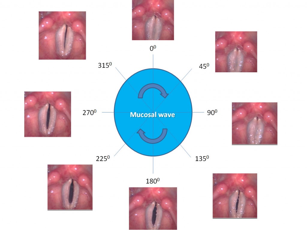
Figure 1
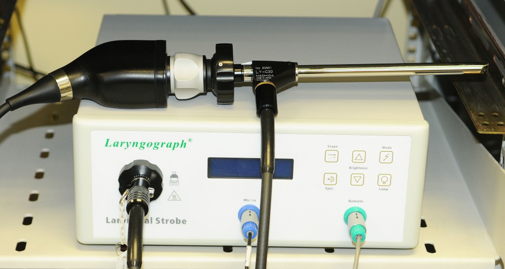
Figure 2
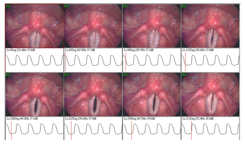
Figure 3
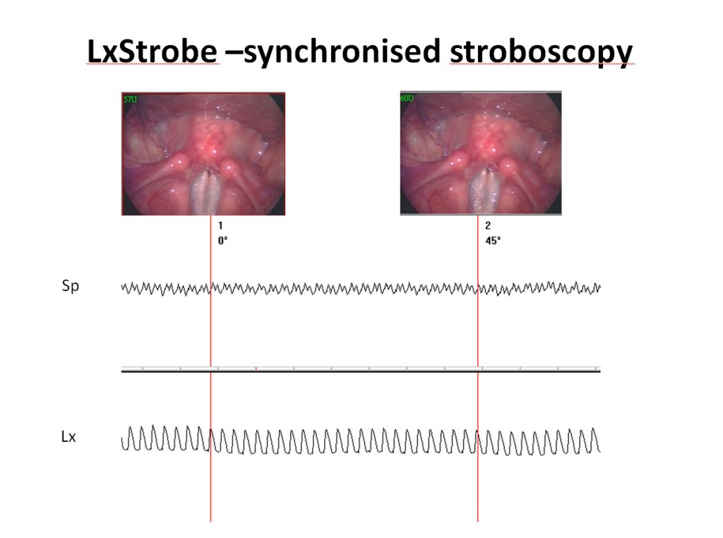
Figure 4